EXPERIMENTATION
FORTH

FORTH
Key Research Facilities, Infrastructure & Equipment
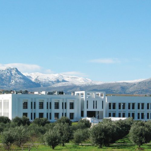
ICS-FORTH maintains state-of-the-art network and computing infrastructures, including clusters of servers (e.g., with GPUs) with extensive computational and storage capabilities. TNL maintains a number of servers, network testbeds, and various spectrum monitoring equipments. IMBB-FORTH is supported by state of the art facilities such as (1) Genomics and postgenomics, (2) Advanced microscopy, (3) Optical tomography, (4) Cell culture, (5) Mouse transgenesis/ mutagenesis, (6) Histology (7) Neurophysiology, and (8) Proteomics.
ICS-FORTH maintains state-of-the-art network and computing infrastructures, including clusters of servers (e.g., with GPUs) with extensive computational and storage capabilities. TNL maintains a number of servers, network testbeds, and various spectrum monitoring equipments. IMBB-FORTH is supported by state of the art facilities such as (1) Genomics and postgenomics, (2) Advanced microscopy, (3) Optical tomography, (4) Cell culture, (5) Mouse transgenesis/ mutagenesis, (6) Histology (7) Neurophysiology, and (8) Proteomics.
Harvard
Key Research Facilities, Infrastructure & Equipment
Dr. Smirnakis lab is equipped with 2-photon microscopes, electrophysiology rigs (both in vitro and in vivo), lasers necessary for optogenetic experiments, the spatial light modulation module.
Dr. Smirnakis lab is equipped with 2-photon microscopes, electrophysiology rigs (both in vitro and in vivo), lasers necessary for optogenetic experiments, the spatial light modulation module.

Harvard

Key Research Facilities, Infrastructure & Equipment
Dr. Smirnakis lab is equipped with 2-photon microscopes, electrophysiology rigs (both in vitro and in vivo), lasers necessary for optogenetic experiments, the spatial light modulation module.
Dr. Smirnakis lab is equipped with 2-photon microscopes, electrophysiology rigs (both in vitro and in vivo), lasers necessary for optogenetic experiments, the spatial light modulation module.
UCLA

Experiments of Tasks
1.1 - 1.2 & 2.1 - 2.3
FORTH
Key Research Facilities, Infrastructure & Equipment
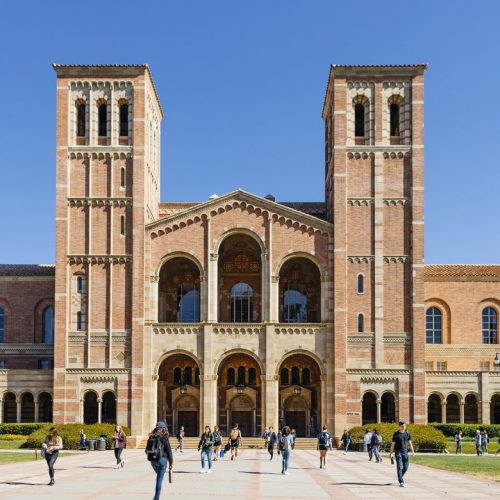
The Department of Neurology at the David Geffen School of Medicine has state-of-the-art facilities for neuroscience research, including an animal facility, facilities neurogenetics and neuroimaging, avanced light microscopy facility, stem cell biology core facility, electron imaging center, behavioral core facility. The Golshani lab is also equipped with 2-photon microscopes and equipment for in vivo and in vitro electrophysiological experiments.
The Department of Neurology at the David Geffen School of Medicine has state-of-the-art facilities for neuroscience research, including an animal facility, facilities neurogenetics and neuroimaging, avanced light microscopy facility, stem cell biology core facility, electron imaging center, behavioral core facility. The Golshani lab is also equipped with 2-photon microscopes and equipment for in vivo and in vitro electrophysiological experiments.

UCLA
Key Research Facilities, Infrastructure & Equipment
The Department of Neurology at the David Geffen School of Medicine has state-of-the-art facilities for neuroscience research, including an animal facility, facilities neurogenetics and neuroimaging, avanced light microscopy facility, stem cell biology core facility, electron imaging center, behavioral core facility. The Golshani lab is also equipped with 2-photon microscopes and equipment for in vivo and in vitro electrophysiological experiments.
The Department of Neurology at the David Geffen School of Medicine has state-of-the-art facilities for neuroscience research, including an animal facility, facilities neurogenetics and neuroimaging, avanced light microscopy facility, stem cell biology core facility, electron imaging center, behavioral core facility. The Golshani lab is also equipped with 2-photon microscopes and equipment for in vivo and in vitro electrophysiological experiments.
UA
Key Research Facilities, Infrastructure & Equipment
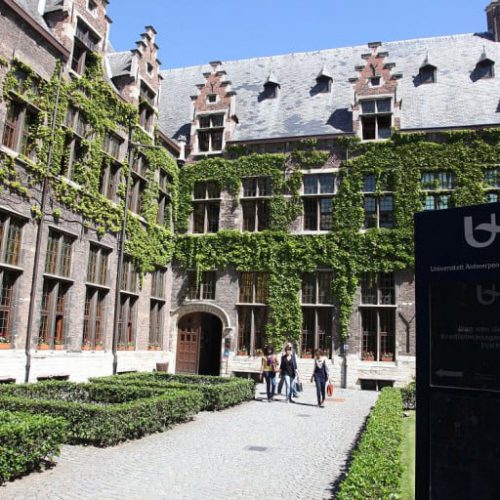
The Bio-Imaging Laboratory is a core in vivo imaging facility and a pioneer in high-resolution small animal MRI neuro-imaging to study learning, neurodegeneration, and neuroplasticity. It houses state of the art pre-clinical MRI scanners including two Pharmascan 7 Tesla MRI systems (Bruker), one BioSpec 9.4 Tesla MRI scanner (Bruker) and one desktop 4.7 Tesla MRI system MRSolution), functional magnetic resonance imaging to identify anatomical regions impacted by released therapeutics in cuprizone and EAE models of inflammatory de- and remyelination models of MS and proton magnetic resonance spectroscopy to profile cortical integrity in treated and non-treated animals. The Bio-Imaging Laboratory has pioneered new imaging paradigms for MRI in small animals including diffusion tensor imaging (DTI)), diffusion kurtosis imaging (DKI), resting state fMRI, ASL-perfusion.
The Bio-Imaging Laboratory is a core in vivo imaging facility and a pioneer in high-resolution small animal MRI neuro-imaging to study learning, neurodegeneration, and neuroplasticity. It houses state of the art pre-clinical MRI scanners including two Pharmascan 7 Tesla MRI systems (Bruker), one BioSpec 9.4 Tesla MRI scanner (Bruker) and one desktop 4.7 Tesla MRI system MRSolution), functional magnetic resonance imaging to identify anatomical regions impacted by released therapeutics in cuprizone and EAE models of inflammatory de- and remyelination models of MS and proton magnetic resonance spectroscopy to profile cortical integrity in treated and non-treated animals. The Bio-Imaging Laboratory has pioneered new imaging paradigms for MRI in small animals including diffusion tensor imaging (DTI)), diffusion kurtosis imaging (DKI), resting state fMRI, ASL-perfusion.


Task 1.1
Experimental data collection for V1 neuronal networks after passive learning, using in vitro calcium
imaging and patch-clamp recording techniques. (Harvard, FORTH)
In this task, mice will be presented with similar visual stimuli repeatedly. Following the presentation, brain
slices from these mice will be taken in order to perform calcium imaging experiments and concurrent patch-clamp
recordings in vitro. To goal of this task is to identify how spontaneous neuronal networks in VI are shaped by the
electrophysiological properties of the neurons participating in the networks and whether this is altered by passive stimulus
presentation.
Task 1.2
Experimental data collection for V1 neuronal networks after active learning using in vitro calcium
imaging and patch-clamp recording techniques. (Harvard, FORTH)
In this task, mice will be trained to discriminate between visual stimuli. Following the training, brain slices
from these mice will be taken in order to perform calcium imaging experiments and concurrent patch-clamp recordings in
vitro. To goal of this task is to identify how spontaneous neuronal networks in VI are shaped by the electrophysiological
properties of the neurons participating in the networks and whether this is altered by training to actively discriminate
between stimuli. Data from this task will further analyzed in WP3.
Task 1.3
Experimental data collection for V1 neuronal networks during passive learning using 2-photon calcium
imaging in vivo. (Harvard, FORTH)
Initially, surgeries will be performed in order
to express the GCamp6s protein in V1 neurons. Following animal recovery, experiments will be performed in
head-restrained animals in order to image activation of neuronal networks during repeated stimulus presentation. This task
will identify how spontaneous neuronal networks in VI are shaped by the electrophysiological properties of the neurons
participating in the networks and whether this is altered by training to actively discriminate between stimuli.
Task 1.4
Experimental data collection for V1 neuronal networks during active learning using 2-photon calcium
imaging in vivo. (Harvard, FORTH)
Initially, surgeries will be performed in order to express the GCamp6s protein in V1 neurons. Following animal recovery,
experiments will be performed in head-restrained animals in order to image activation of neuronal networks during
stimulus discrimination training.
Task 1.5
SLM causal interrogation. (Harvard, FORTH)
In this task, putative causal relationships of firing between neuronal sub-networks will be identified
statistically and then experimentally probed using state-of-the-art spatial light modulation (SLM) technology in
conjunction with channelrhodopsin (C1V1) expression. One important question is whether activation of “bridge” neurons
(neurons with multiple functional connectivity links to different sub-networks) recruits the sub-networks they connect.
Task 2.1
Experimental data collection for spontaneous PFC neuronal networks in vitro. (FORTH, Harvard)
In this
task, PFC brain slices will be prepared from adult mice in order to perform concurrent calcium imaging experiments,
following incubation with fura-2AM, concurrently with patch-clamp recordings in order to identify spontaneously
activated neuronal networks in the PFC. The goal is to generate data for analysis in WP3.
Task 2.2
Experimental data collection for PFC networks following passive learning in vitro. (FORTH, Harvard)
Initially, surgeries will be performed in order to express the GCamp6s protein in V1 neurons. Following animal recovery,
experiments will be performed in head-restrained animals in order to image activation of neuronal networks during
stimulus discrimination training.
Task 2.3
Experimental data collection for PFC networks following active learning, using in vitro calcium imaging
and/or patch-clamp recordings.
(FORTH, Harvard)
In this task, putative causal relationships of firing between neuronal sub-networks will be identified
statistically and then experimentally probed using state-of-the-art spatial light modulation (SLM) technology in
conjunction with channelrhodopsin (C1V1) expression. One important question is whether activation of “bridge” neurons
(neurons with multiple functional connectivity links to different sub-networks) recruits the sub-networks they connect.
Task 2.4
Experimental data collection in vivo for PFC networks during the spontaneous alternation task. (FORTH, UCLA)
Initially, surgeries will be performed in order to express the GCamp6s protein in PFC neurons. A second surgery
will be performed to place the miniscope onto the mouse head. Following animal recovery, experiments will be performed
in freely-moving animals in order to image activation of neuronal networks during spontaneous alternation procedure in
the Y-maze. This task will generate data on neuronal network activation in the PFC during phases of the spontaneous
alternation task that will be analyzed in WP3.
Task 2.5
Experimental data collection in vivo for PFC networks during passive learning. (FORTH,
UCLA)
Animal
surgeries will be performed as outlined in task 2.4. Following animal recovery, experiments will be performed in
freely-moving animals in order to image activation of neuronal networks during training in the delayed alternation
procedure in the T-maze. This task will generate data on neuronal network activation in the PFC during different days and
different phases of the delayed alternation task that will be analyzed in WP3.
Global network monitoring (UA)
Performing rsfMRI for global, whole-brain network analysis: The
high field functional magnetic resonance imaging (fMRI) offers great opportunities to study this system at
a global, whole-brain network-level, while the resting state functional magnetic resonance imaging
(rsfMRI) allows the visualization of the functional connectivity (FC) between different areas in the brain.
The Antwerp Bio-Imaging Lab (BIL) established itself as a major international player in this field by
publishing the first papers in mice. Soon after several groups followed this interesting approach.
A key advantage of performing MRI in small animal models is a possibility for combining such a global
network approach with many other neuroscientific tools (i.e. genetics) allowing to manipulate the system at
very specific sites. Moreover, MRI can be used in parallel with other readouts such as electrophysiology,
and optical imaging, to provide information at multiple spatiotemporal scales.
Optogenetic functional magnetic resonance imaging (UA)
OfMri is an innovative technique that,
combining the precision of optogenetic manipulation and the global coverage of fMRI, allows to examine
the FC of specific neural networks at the whole-brain level. In this case, fMRI helps to determine
connectivity changes in response to optogenetic manipulation without electromagnetic interference with
radio-frequency (RF) signal. Notably, ofMRI in rodents could be fundamental in translating neural
networks knowledge obtained with animal studies toward clinics in humans.
